| |
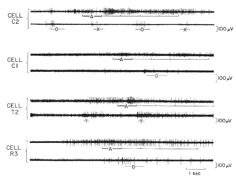
Figure 8: Cells facilitated during affective defense 50% more than by any control manipulation | Page 23 |
Cell C2 from the midbrain central gray is shown firing at maximal rate during affective defense (A) and also responding to opening of the partition (O) and closing of the partition (X) which separated the cats. A smaller cell may also be seen firing before and after the affective defense. Changes in spike amplitude of cell C2 were due to tissue movement. Cell C1 from the midbrain central gray is shown responding at its maximal rate during and after affective defense (A) and at a lesser rate during dropping of the cat (D). Cells R3 and T2, each located just medial to the mamillo-thalamic tract, are shown firing at maximal rates during affective defense (A) and at lesser rates during tapping on the partition separating the cats (T) and dropping of cat (D).
 |
 |
 |